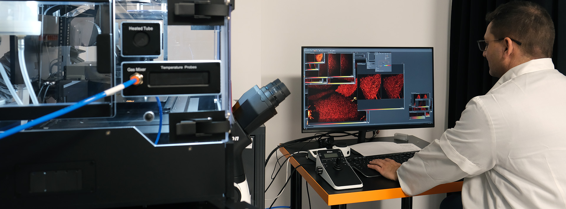
In TIRF (Total Internal Reflection Fluorescence) microscopy the excitation beam strikes the sample-coverslip interface at a sufficiently high angle, termed the critical angle. Although the light basically doesn't passes into the sample, it generates a highly restricted electromagnetic field adjacent to the interface. This evanescent field is identical in frequency to the excitation beam and it's intensity decays exponentially with distance from the interface. The fluorescent excitation can be induced in a very thin (150 nm) optical section, which immediately adjacent to the sample-coverslip interface. Due to the negligible background the method provides images with excellent contrast with the ability of good time resolution. This method is ideal for single molecule detection in a thin optical section, typically in membranes.
- Microscope: Olympus IX81S1F-ZDC2
- Camera: Hamamatsu C9300
- Lasers: 491nm, 640nm
- Objectives: 10x, 20x, 60x, 100x
