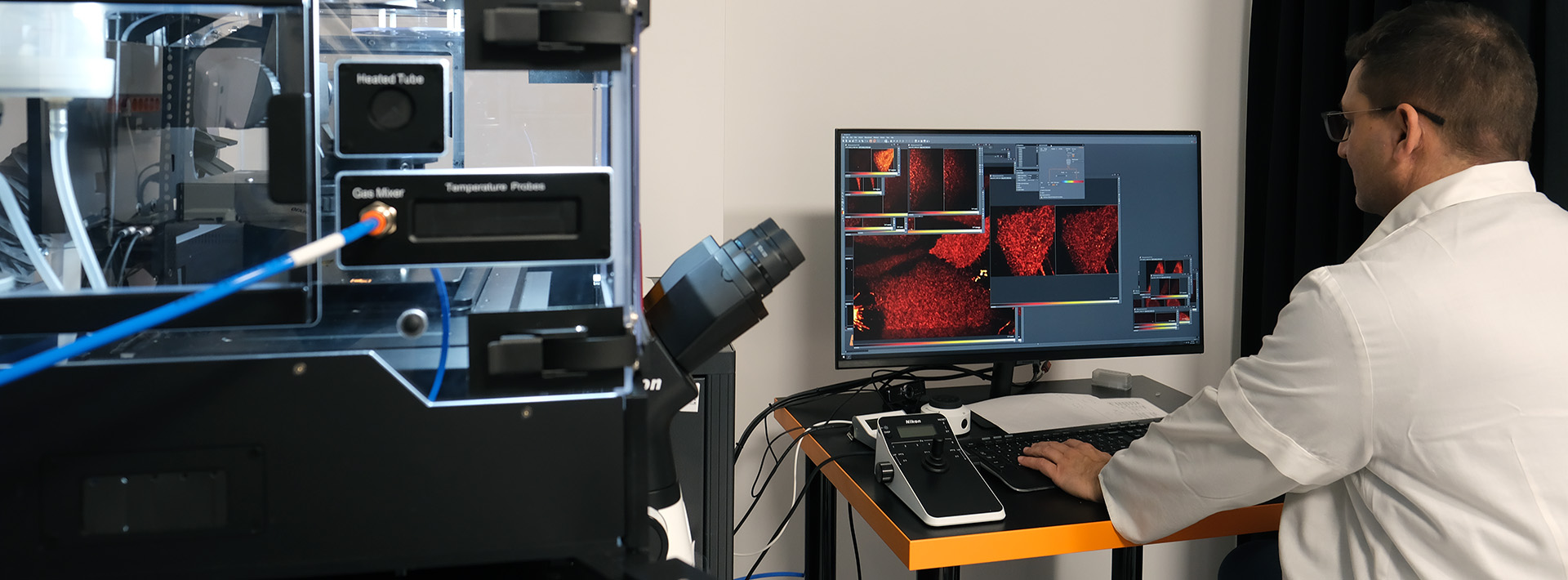
In the case of “Structured Illumination Microscopy” (SIM), an increase in resolution is achieved by modulating the excitation beam so that the excitation illumination pattern varies with location. When taking a SIM image, the pattern is illuminated from different directions and in different phases with the given and known pattern. We can roughly double the resolution in the x-y direction by using the Fourier analysis on the two-dimensional measured images. With the method approx. 33 fps imaging is available, making it suitable for dynamic scanning. The microscope available at the Nano-Bio-Imaging Core Facility is also suitable for live-cell studies as it is equipped with a CO2 and temperature incubator too.
- Microscope: Zeiss Elyra S.1
- Objectives: Plan-Neofluar 10x/0.30, Plan-APO 40x/1.4 Oil, Plan-Apochromat 63x/1.40 Oil, Alpha Plan-APO 100x/1.46 Oil
- Lasers: 405 nm, 488 nm, 561 nm és 642nm
- Filters:405nm excitation (MBS 405 + EF BP 420-480 / LP 750), 488nm excitation (MBS 488 + EF BP 495-550 / LP 750), 561nm excitation (MBS 561 + EF BP 570-620 / LP 750), 642nm excitation (MBS 642 + EF LP 655)
- Camera: Andor iXon EMCCD
