Data
Official data in SubjectManager for the following academic year: 2024-2025
Course director
-
Bugyi Beáta
associate professor,
Department of Medical Biology and Central Electron Microscope Laboratory -
Number of hours/semester
lectures: 12 hours
practices: 0 hours
seminars: 0 hours
total of: 12 hours
Subject data
- Code of subject: OPF-MKA-T
- 1 kredit
- Pharmacy
- Optional modul
- spring
-
Course headcount limitations
min. 5 – max. 15
Available as Campus course for . Campus-karok: GYTK TTK
Topic
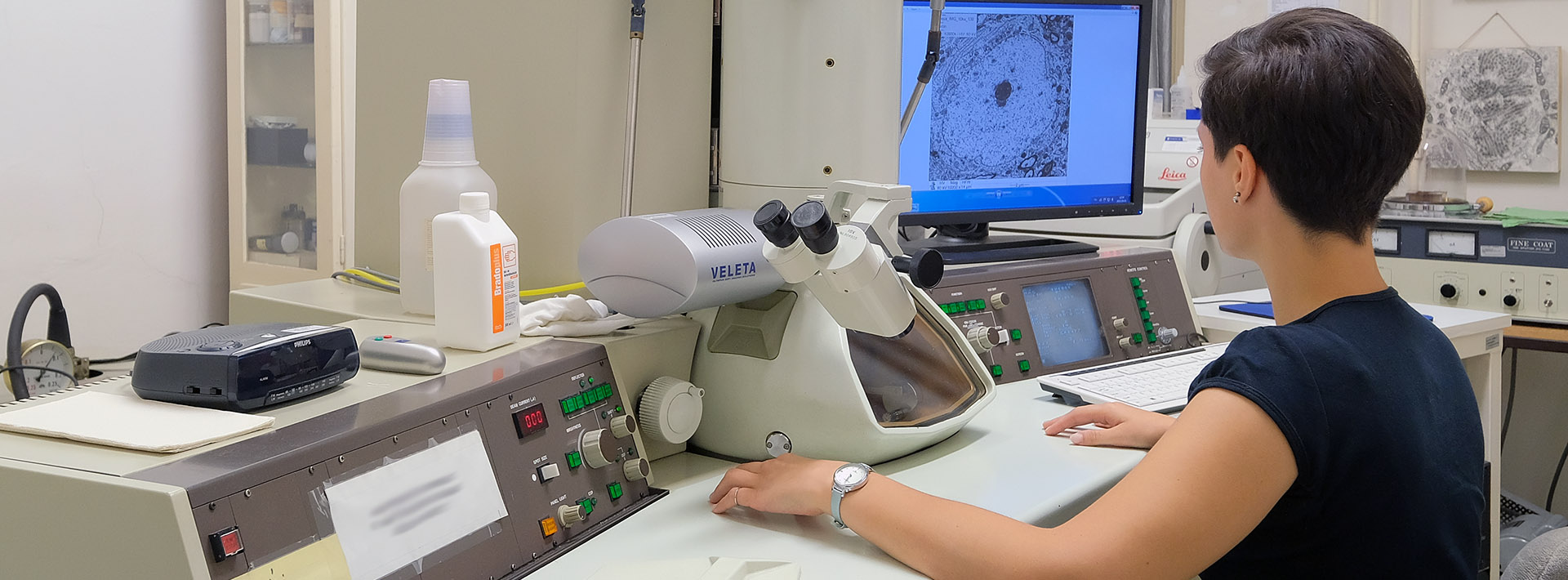
Modern microscopic methods used in biomedical research, as well as the principles and practice of related digital image processing and analysis, are presented. Students can acquire knowledge related to microscopic imaging methods and image analysis by solving practical problems, analyzing case studies, and doing independent computer work (image analysis software, e.g. ImageJ, Fiji, Imaris, etc.). Strong emphasis is laid on hands-on skills. The course supports practical skills that help research or Students’ research (TDK) work and can contribute to a better understanding of the corresponding topics of compulsory subjects (e.g. molecular cell biology, cell biology, histology, anatomy).
Module 1 - introduction: Basic principles of microscopic imaging methods.
Module 2 - interactive sessions: Practical aspects of microscopic imaging methods.
Module 3 - interactive sessions: Basics of digital image processing and analysis.
Lectures
- 1.
Basic principles of microscopic imaging methods used in biomedical research.
- Bugyi Beáta - 2.
Basic principles of microscopic imaging methods used in biomedical research.
- Leipoldné Vig Andrea Teréz - 3.
Light microscopy. Fluorescence microscopy. How to build a light microscope.
- Bugyi Beáta - 4.
Light microscopy. Fluorescence microscopy. How to build a light microscope.
- Bugyi Beáta - 5.
Electron microscopy.
- Bugyi Beáta - 6.
Cryo-electron microscopy.
- Bugyi Beáta - 7.
Image analysis 1.
- Bugyi Beáta - 8.
Image analysis 2.
- Bugyi Beáta - 9.
Image analysis 3.
- Gaszler Péter - 10.
Image analysis 4.
- Gaszler Péter - 11.
Workshop. Students' projects and presentations.
- Bugyi Beáta, Szütsné Tóth Mónika Ágnes - 12.
Workshop. Students' projects and presentations.
- Bugyi Beáta, Szütsné Tóth Mónika Ágnes
Practices
Seminars
Reading material
Obligatory literature
Literature developed by the Department
Can be found on MS Teams group of the course.
Notes
Recommended literature
John C. Russ Introduction to Image Processing and Analysis ISBN-13: 978-0849370731
Toennies, Klaus D. Guide to Medical Image Analysis ISBN: 978-1-4471-7320-5
Chris Solomon & Toby Breckon Fundamentals of Digital Image Processing ISBN-13: 978-0470844731
Conditions for acceptance of the semester
A maximum of 25% absence is allowed.
Mid-term exams
Grading policy
The grade is based on the result of
a written test,
or short (5-10 minutes) presentations of Students’ projects.
Making up for missed classes
The opportunity to make up for absence can be discussed with the course leader.
Exam topics/questions
No exam is scheduled in the exam period.
