Adatok
A Tantárgybejelentőben megadott hivatalos adatok az alábbi tanévre: 2019-2020
Tantárgyfelelős
-
Dr. Hajnalka Gabriella ÁBRAHÁM
associate professor,
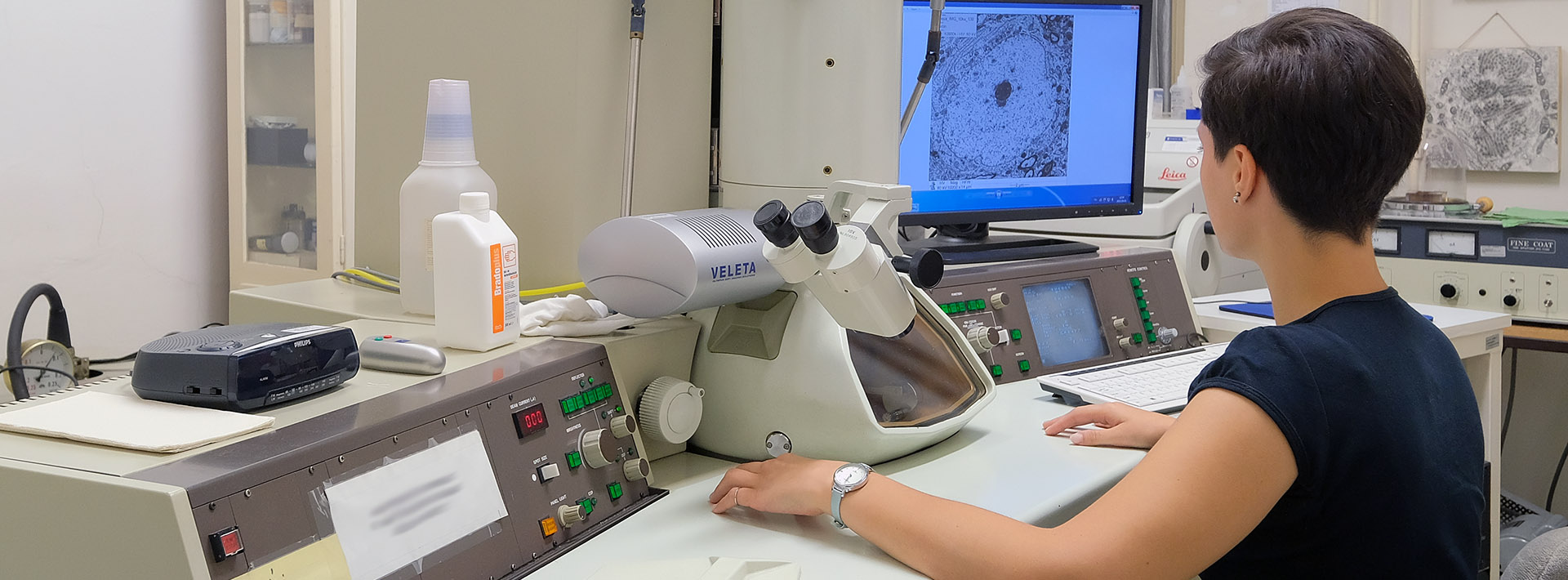
Department of Medical Biology and Central Electron Microscope Laboratory
Óraszámok/félév
előadás: 24 óra
gyakorlat: 0 óra
szeminárium: 0 óra
összesen: 24 óra
Tárgyadatok
- Kód: OSF-EMA-T
- 2 kredit
- Dentistry
- Optional modul
- spring
Nincs
Vizsgakurzus:Nem
Kurzus létszámkorlát
min. 5 fő – max. 25 fő
Tematika
Methods of electron microscopic fixation, embedding and sectioning and the use of the electron microscope. Demonstration of the subcellular elements in details and of a few tissues and organs. Demonstration of the electron microscopic methods used in the biological research. The use of electron microscopic methods in the clinical practice.
The basic principles of electron microscopy will be discussed and we provide information about the mode and the use of electron microscopy in the basic research and in the clinical practice.
Előadások
- 1. Purpose and indication of the electron microscopic examination. The role of fixation in tissue preservation, the recognition of cells, cellular organelles. - Dr. Rimayné Dr. Ábrahám Hajnalka Gabriella
- 2. Most frequent failures in the electronmicroscopic practice - Dr. Seress László Antal
- 3. Optimal fixation for electron microscopy. Composition of fixatives for different tissue samples. - Dr. Seress László Antal
- 4. Samples taken by autopsy or biopsy. Perfusion of experimental animals for electron microscopy. Electron microscopy of formaldehyde fixed and paraffin embedded materials. - Dr. Seress László Antal
- 5. The ultrastructure of the cell I - Dr. Seress László Antal
- 6. The ultrastructure of the cell II - Dr. Seress László Antal
- 7. The ultrastructure of the cell III - Dr. Seress László Antal
- 8. The ultrastructure of the cell IV - Dr. Seress László Antal
- 9. The ultrastructure of the cell V - Dr. Rimayné Dr. Ábrahám Hajnalka Gabriella
- 10. The ultrastructure of the cells VI. Test. - Dr. Rimayné Dr. Ábrahám Hajnalka Gabriella
- 11. Ultrastructures of neurons (axon, dendrite, synapses) - Dr. Seress László Antal
- 12. Ultrastructure of glial cells - Dr. Seress László Antal
- 13. The ultrastructure of kidney - Dr. Rimayné Dr. Ábrahám Hajnalka Gabriella
- 14. The ultrastructure of the liver - Dr. Rimayné Dr. Ábrahám Hajnalka Gabriella
- 15. Ultrastructure of the muscle. - Dr. Rimayné Dr. Ábrahám Hajnalka Gabriella
- 16. Necrosis and apoptosis. - Dr. Rimayné Dr. Ábrahám Hajnalka Gabriella
- 17. Combined light and electron microscopic methods, such as Golgi/EM, Timm/EM, immunocytochemistry/EM. - Dr. Seress László Antal
- 18. Combination of degeneration and axon transport methods with electron microscopy. - Dr. Seress László Antal
- 19. Ultrastructure of bacteria and viruses. - Dr. Rimayné Dr. Ábrahám Hajnalka Gabriella
- 20. Cytoskeleton - Dr. Seress László Antal
- 21. Intercellular connections - Dr. Seress László Antal
- 22. The use of EM in biological research I - Dr. Seress László Antal
- 23. The use of EM in biological research II - Dr. Seress László Antal
- 24. Exam - Dr. Seress László Antal
Gyakorlatok
Szemináriumok
A tananyag elsajátításához szükséges segédanyagok
Kötelező irodalom
Saját oktatási anyag
Jegyzet
Ajánlott irodalom
Suggested only:
Dr. Szabolcs Virágh: Ultrastructural Pathology and Diagnostic Electron Microscopy
Leon Weiss: Histology. Cell and Tissue Biology
A félév elfogadásának feltételei
Participation on 80% of the classes. Oral examination with analysis of 3 electron microscopic photographs
Félévközi ellenőrzések
No possibility
Távolmaradás pótlásának lehetőségei
No possibility
Vizsgakérdések
Exam topics are the same as lecture topics